- Applications
- Life Science Applications, Micro-CT Systems Applications
- Micro-CT for Zoology Research
- Applications
- Life Science Applications, Micro-CT Systems Applications
- Micro-CT for Zoology Research
Micro-CT for Zoology Research


BRUKER SKYSCAN X-ray tomography instruments apply to all forms of life. The power of 3D nondestructive imaging has revolutionized the way zoologists conduct integrative and comparative biology research.
- High sensitivity to different tissue types
- Superior resolution for resolving subtle differences in morphometry
- Comprehensive visualization tools to produce stunning images that are sure to attract attention
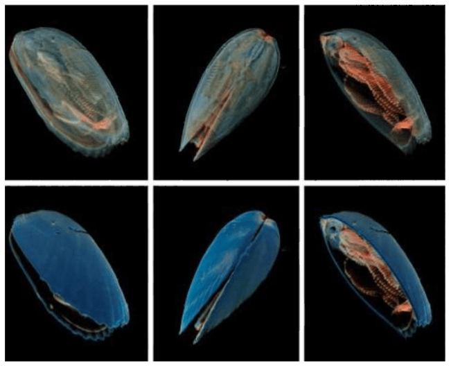
Comparative biology
The SkyScan visualization programs DATAVIEWER and CTVOX create beautifully rendered images and movies of specimens, allow for complete data manipulation, and side-by-side comparison in 3D space.
Model biomechanics & functional biology
High quality micro-CT images are the first step in modeling biomechanics and functional biology.
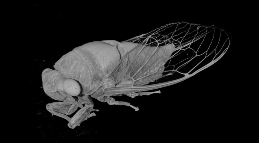
Entomology
Our comprehensive method notes provide a blueprint to extract the most details out of samples which inherently do not provide natural X-ray contrast. It is possible through the use of prescribed protocols to obtain detailed images at ultra-high resolution. Through the use of the SkyScan 3D Suite visualization tools, breath-taking rendering can be created from your samples.

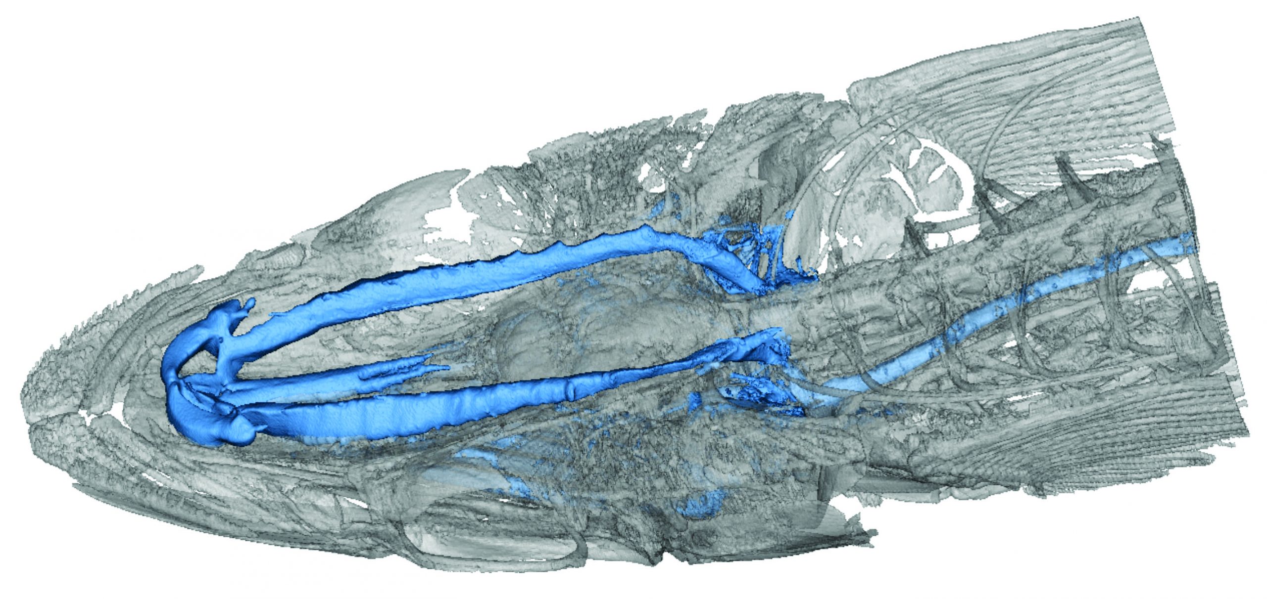
Micro-CT for vertebrates
X-ray computed tomography for vertebrates does not mean being restricted to skeletal anatomy. Through the use of contrast enhancing agents and chemical drying techniques, a full range of tissue information can be extracted from the micro-CT images.
Related Articles
Micro-CT Imaging of Avian Skulls
Figure 1: Images of a Chukar Partridge and a Pheasant 1,2 With nearly 11,000 species of living birds comprising a vast spectrum of shapes, colors,
Seth Hogg, PhD
September 28, 2021
Applications of Micro-CT for Avian Studies
Micro-CT’s non-destructive, high resolution 3D imaging makes it one of the most important tools for morphological avian research as well as biomimetic studies. One area
Ann Bagnell
September 28, 2021
Applications of Micro-CT for Natural History Specimens
Syllis gracilis (a species of marine bioluminescent polychaete worms): false-colour volume rendering of virtually dissected proventricle. The length of the proventricle and the number of
Ann Bagnell
July 29, 2021
Micro-CT Imaging of a Triggerfish
Figure 1: Micro-CT volumetric rendering of a triggerfish imaged using the SkyScan 1273 To best match the wide range of sample sizes encountered in cataloging
Seth Hogg, PhD
July 29, 2021
Not sure about the best instrument for your application?
Do you want to see micro-CT results of your sample?
Our specialists are happy to help.
Do you want to see micro-CT results of your sample?
Our specialists are happy to help.
Not sure about the best instrument for your application?
Do you want to see micro-CT results of your sample?
Our specialists are happy to help.
Do you want to see micro-CT results of your sample?
Our specialists are happy to help.